Doctors have successfully diagnosed a rare congenital condition in a four-week-old infant using imaging techniques alone, avoiding the need for invasive surgery and highlighting advancements in paediatric care.
Rare condition detected early
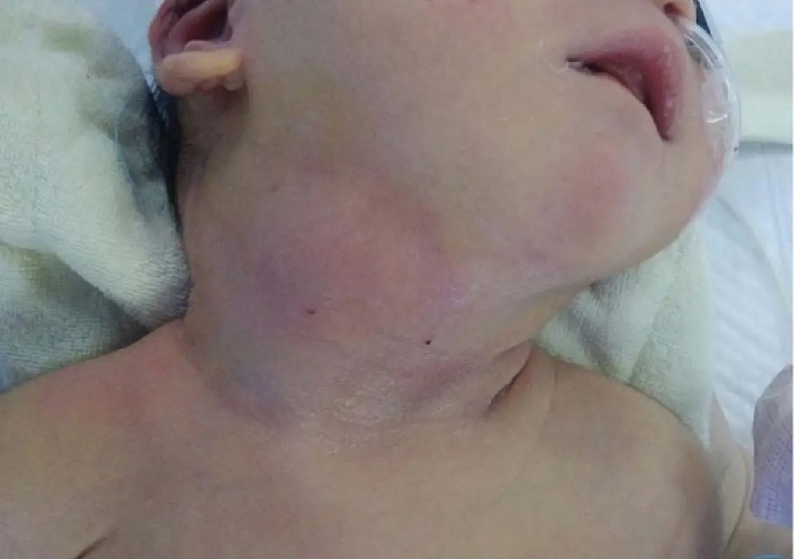
The infant presented with swelling on the left side of the neck, first noticed at three weeks of age. The condition was identified as ectopic cervical thymus (ECT), a rare anomaly caused by incomplete movement of thymus tissue during fetal development.
Due to its rarity, ECT is often mistaken for other serious conditions, including malignant neck tumours.
Imaging helps avoid surgery
Doctors used ultrasonography and magnetic resonance imaging (MRI) to evaluate the swelling. The scans revealed features identical to normal thymus tissue, including a characteristic internal pattern and similar signal intensity.
Importantly, there was no compression of nearby airways or blood vessels, supporting a non-invasive diagnosis.
Conservative treatment proves effective
Based on imaging findings, doctors opted for conservative management instead of surgical removal, which is commonly performed in such cases to rule out cancer.
The infant has since shown gradual improvement, with the swelling reducing over time.
Significance for paediatric care
This case highlights the importance of accurate imaging and careful comparison with normal anatomical structures in diagnosing rare conditions.
Experts note that such approaches can help avoid unnecessary surgical procedures, reduce risks for infants, and promote safer treatment pathways.
A step forward in diagnosis
The successful non-invasive diagnosis demonstrates how modern imaging techniques can improve clinical decision-making, particularly in rare paediatric conditions where traditional approaches often involve surgery.